

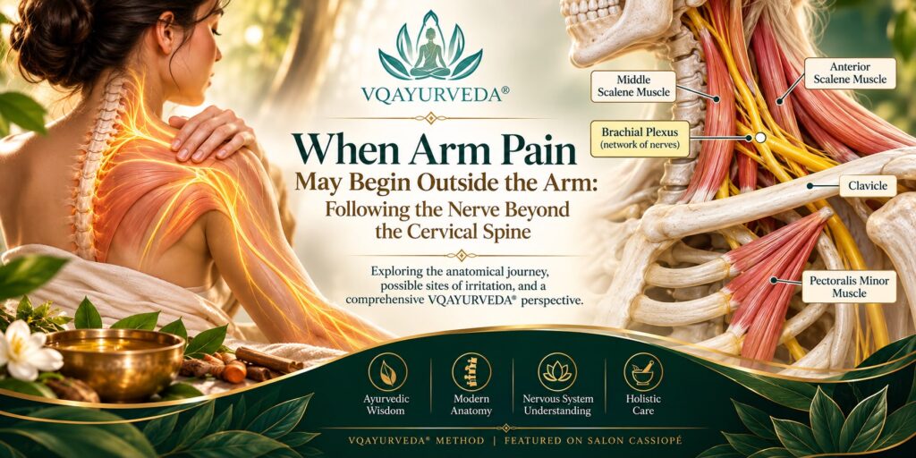
Pain naturally draws our attention to the place where we feel it.
When the hand tingles, we focus on the hand. When the forearm aches, we massage the forearm. When the shoulder feels tight, we stretch the shoulder.
This makes intuitive sense. But neurologically, the location of a symptom does not always identify the location where the physiological disturbance begins.
The nerves supplying the upper limb travel a considerable distance. Along their journey from the spinal region toward the shoulder, arm and hand, they pass through a complex anatomical environment of muscles, fascia, bones, joints and blood vessels.
This raises an important question:
What if recurring arm pain, tingling or numbness is not always an arm problem — and does not necessarily begin inside the cervical spine?
The Journey From the Spine to the Arm
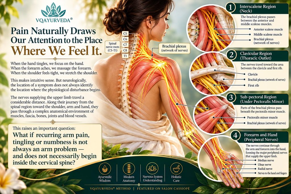
The neural pathways supplying much of the upper limb begin with spinal nerve roots from the lower cervical and upper thoracic regions. C5, C6, C7, C8 and T1 The anterior rami of C5, C6, C7, C8 and T1 primarily form a complex network of nerves known as the brachial plexus.
The brachial plexus is not simply one nerve running from the neck into the arm. It is an organized network in which nerve fibres combine, divide and reorganize before ultimately contributing to the major peripheral nerves supplying the upper limb.
To reach the arm, these neural structures must travel through several relatively confined anatomical regions. Near the neck, the roots and trunks of the brachial plexus pass through the interscalene triangle, a space bordered primarily by the anterior scalene, middle scalene and first rib. They then travel toward the region between the clavicle and first rib, before continuing toward the shoulder.
Farther along this pathway, portions of the brachial plexus pass beneath the pectoralis minor muscle before the major peripheral nerves continue into the arm. This anatomical journey reveals something important:
Once a nerve has left the spine, its relationship with surrounding structures continues. The Spine Is Only One Possible Location Radiating arm pain, tingling or numbness is frequently associated with the idea of a “pinched nerve” in the neck. And sometimes that is exactly where the problem lies.
A cervical nerve root can become irritated by several mechanisms, including inflammatory processes, degenerative changes or narrowing around the space through which the nerve root exits. But neural symptoms can also arise farther along the pathway.
One recognized example is neurogenic thoracic outlet syndrome, in which elements of the brachial plexus may be affected as they travel from the neck toward the upper limb.
The region beneath the pectoralis minor has also been investigated as another potential location of neural compression or irritation in some individuals.
Depending upon the structures involved, symptoms may include pain, tingling, numbness, altered sensation, heaviness or weakness affecting portions of the shoulder, arm or hand.
However, this requires an important qualification: A tight scalene or pectoralis minor muscle does not automatically mean that a nerve is compressed.
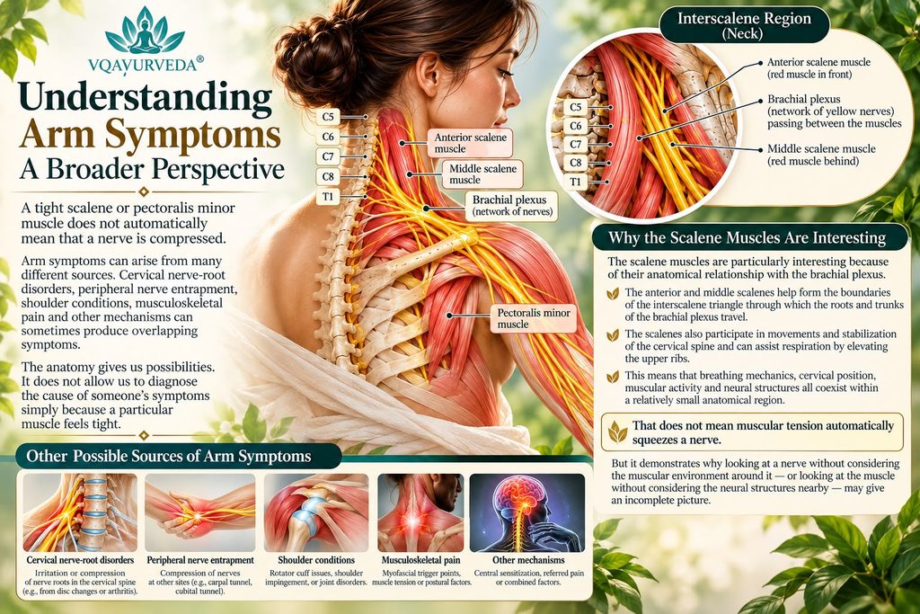
Arm symptoms can arise from many different sources. Cervical nerve-root disorders, peripheral nerve entrapment, shoulder conditions, musculoskeletal pain and other mechanisms can sometimes produce overlapping symptoms.
The anatomy gives us possibilities. It does not allow us to diagnose the cause of someone’s symptoms simply because a particular muscle feels tight.
Why the Scalene Muscles Are Interesting
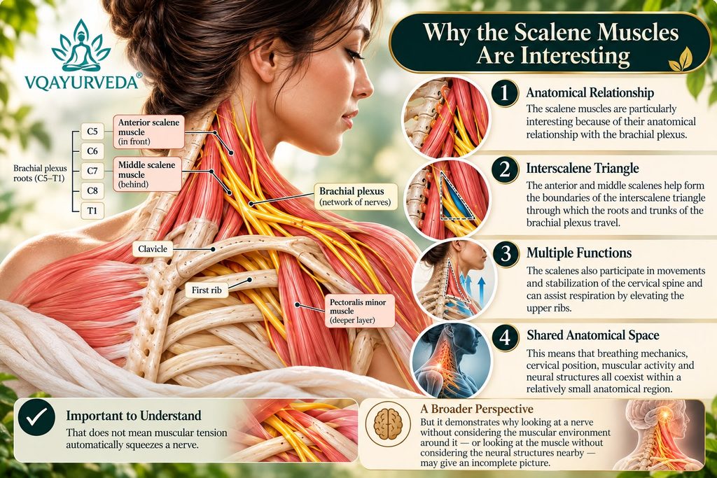
The scalene muscles are particularly interesting because of their anatomical relationship with the brachial plexus.
The anterior and middle scalenes help form the boundaries of the interscalene triangle through which the roots and trunks of the brachial plexus travel. The scalenes also participate in movements and stabilization of the cervical spine and can assist respiration by elevating the upper ribs.
This means that breathing mechanics, cervical position, muscular activity and neural structures all coexist within a relatively small anatomical region.
That does not mean muscular tension automatically squeezes a nerve. But it demonstrates why looking at a nerve without considering the muscular environment around it — or looking at the muscle without considering the neural structures nearby — may give an incomplete picture.
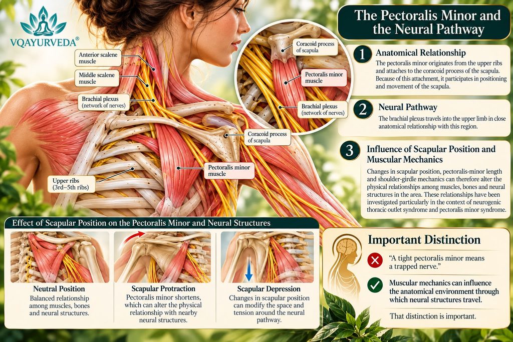
The Pectoralis Minor and the Shoulder Girdle. The same principle becomes interesting farther along the neural pathway. The pectoralis minor originates from the upper ribs and attaches to the coracoid process of the scapula.
Because of this attachment, it participates in positioning and movement of the scapula.
The brachial plexus travels into the upper limb in close anatomical relationship with this region.
Changes in scapular position, pectoralis-minor length and shoulder-girdle mechanics can therefore alter the physical relationships among muscles, bones and neural structures in the area. These relationships have been investigated particularly in the context of neurogenic thoracic outlet syndrome and pectoralis minor syndrome.
Again, the important point is not: “A tight pectoralis minor means a trapped nerve.”
The more scientifically appropriate observation is:
Muscular mechanics can influence the anatomical environment through which neural structures travel. That distinction is important.
A Nerve Is Living, Responsive Tissue
We sometimes imagine nerves almost like electrical wires fixed inside the body.
Physiology is considerably more dynamic. Peripheral nerves must accommodate normal body movement while maintaining their ability to conduct neural signals.
When we move the neck, shoulder, elbow, wrist or fingers, the mechanical relationships between nerves and surrounding tissues change.
At the same time, sensory information is constantly travelling toward the spinal cord and brain.
The nervous system receives information from the skin, muscles, joints and other tissues, integrates those signals and continually adjusts motor activity.
This means that muscle and nerve are not operating independently. They are participating in an ongoing neuromuscular conversation.
Why Does a Muscle Become Tight?
Muscular tightness can have many explanations.
A muscle may be shortened, overloaded or fatigued. Movement patterns may repeatedly recruit it. Joint mechanics can influence its activity.
But muscular tension can also occur as part of a protective response.
When the nervous system interprets sensory input as potentially threatening or painful, motor output may change. Increased muscular activity can sometimes help stabilize or protect a sensitive region.
This is often described as muscle guarding.
A simplified cycle might therefore look like this:
Irritation → sensory signaling → pain or sensitivity → protective muscular activity → altered movement → continued sensory input
The relationship can become bidirectional.
Muscular tension may influence the mechanical environment around neural structures, while neural sensitivity may influence muscular activity.
Suddenly the question becomes more interesting than simply:
“Which muscle is tight?”
Why More Stretching Is Not Always the Answer
Stretching is not inherently problematic. Neither are strengthening, exercise or massage.
When muscular shortening or reduced mobility is contributing to discomfort, appropriately selected movement and stretching can be useful. But treatment should correspond to physiology.
If neural sensitivity is contributing to someone’s symptoms, repeatedly forcing a painful stretch or aggressively pressing into an uncomfortable region may simply reproduce the sensory stimulus rather than explain why it is occurring.
This does not mean that a sensitive nerve should never move. Normal neural tissue is designed to accommodate movement.
The distinction is between appropriate movement and simply forcing a structure because it feels tight. Instead of repeatedly asking:
“How can I stretch this tighter?”
another question becomes possible: “Why is the body maintaining tension here?”And beyond that:
“What is happening along the pathway of the nerve?”
From the Painful Point to the Therapeutic Environment
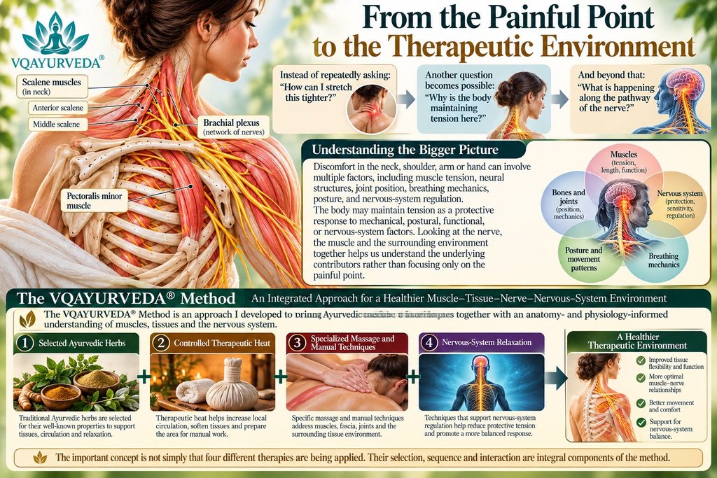
This broader perspective is particularly relevant to the VQAYURVEDA® Method, an approach I developed to bring Ayurvedic therapeutic principles together with an anatomy- and physiology-informed understanding of muscles, tissues and the nervous system.
The method does not look exclusively at the location where discomfort is perceived. It considers the broader muscle–tissue–nerve–nervous-system environment surrounding the problem.
The VQAYURVEDA® Method integrates:
Selected Ayurvedic herbs + controlled therapeutic heat + specialized massage and manual techniques + nervous-system relaxation. The important concept is not simply that four different therapies are being applied.
Their selection, sequence and interaction are integral components of the method.
Ayurveda and the Therapeutic Environment
Traditional Ayurveda has long recognized therapeutic approaches involving oil, warmth and touch. Snehana therapeutic oleation, Snehana, therapeutic oleation, and Swedana, therapeutic heat or sudation, are established concepts within Ayurvedic practice and are particularly relevant to traditional approaches involving Vata.
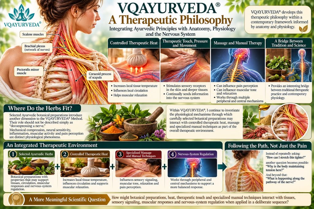
VQAYURVEDA® develops this therapeutic philosophy within a contemporary framework informed by anatomy and physiology.
From a modern physiological perspective, controlled superficial heat can increase local tissue temperature and influence local circulation and muscular relaxation.
Therapeutic touch, pressure and movement stimulate sensory receptors in the skin and deeper tissues, continually sending information into the nervous system.
Massage and manual therapy can also influence pain perception, muscular tone and relaxation through multiple peripheral and central mechanisms.
This provides an interesting bridge between traditional therapeutic practice and contemporary physiology.
Where Do the Herbs Fit?
Selected Ayurvedic botanical preparations introduce another dimension to the VQAYURVEDA® Method.
Their role should not be described simply as “decompressing a nerve.”
Mechanical compression, neural sensitivity, inflammation, muscular activity and pain perception are distinct physiological phenomena.
Within VQAYURVEDA®, I continue to investigate the physiological mechanisms through which carefully selected botanical preparations may interact with controlled therapeutic heat, massage and specialized manual techniques as part of the overall therapeutic environment.
The research question is therefore broader than:
“Which herb treats a nerve?” It becomes:
How might botanical preparations, heat, therapeutic touch and specialized manual techniques interact with tissues, sensory signaling, muscular responses and nervous-system regulation when applied in a deliberate sequence?
That is a much more meaningful scientific question.
Following the Path, Not Just the Pain
The anatomy of the brachial plexus provides a beautiful example of why the human body cannot always be understood as a collection of isolated parts.
A sensation experienced in the fingers may involve neural structures located much farther upstream.
Arm discomfort can relate to the shoulder girdle.
Shoulder mechanics interact with the scapula, clavicle and ribs.
Muscles influence movement and positioning.
Neural structures travel among those muscles and bones.
Sensory information continuously returns to the spinal cord and brain.
And the nervous system continually adjusts muscular activity in response.
The entire system is communicating.
Therefore, when symptoms repeatedly appear in the shoulder, arm or hand, the most interesting question may not always be:
“Where does it hurt?” It may be:
“What pathway leads to where it hurts — and what is happening along that pathway?”
Sometimes the primary issue may be muscular.
Sometimes it may involve a cervical nerve root.
Sometimes neural structures farther from the spine may be involved.
Sometimes altered movement and muscular guarding may contribute.
And sometimes several mechanisms may interact.
Understanding those relationships — rather than automatically chasing the location of pain — is a fundamental principle guiding the
VQAYURVEDA® Method.
VQAYURVEDA® — Where Ayurvedic science meets modern anatomy, physiology, and the nervous system.
VQAYURVEDA® Method | Featured on Salon Cassiopé